
Cellulogia: Structural Skin Condition Explained With Clinical Precision
Cellulogia refers to a structural modification of subcutaneous tissue that alters skin surface morphology. Dermatological literature uses the term cellulogia to describe what is commonly labeled as cellulite. Authoritative medical sources such as Mayo Clinic and American Academy of Dermatology classify cellulogia as a non-disease cosmetic condition. Cellulogia involves adipose tissue, fibrous septae, dermal thickness, and microcirculation. Cellulogia does not indicate inflammation, infection, or metabolic pathology.
Cellulogia presents as surface depressions, nodules, and uneven skin texture. Cellulogia most frequently appears on thighs, buttocks, hips, and lower abdomen. Cellulogia affects appearance rather than physiological function.
Structural Anatomy Responsible for Cellulogia
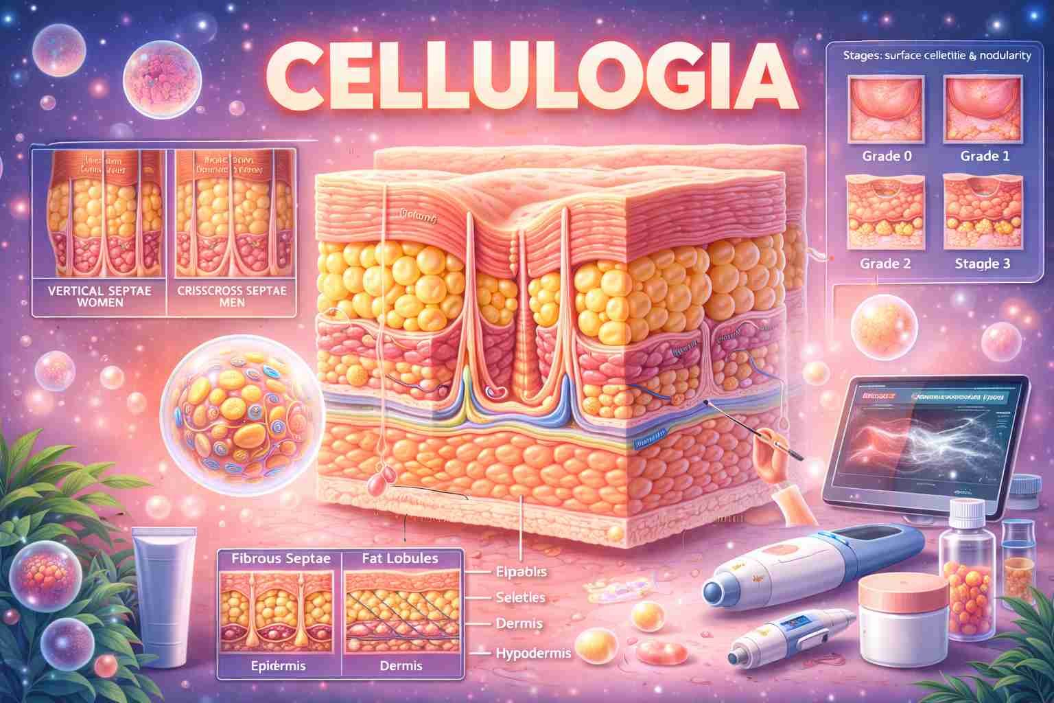
To understand cellulogia, subcutaneous anatomy provides the foundation. Human skin consists of three primary layers: epidermis, dermis, and hypodermis. The hypodermis contains adipocytes grouped into lobules. Fibrous septae connect the dermis to underlying fascia.
Cellulogia forms when adipose lobules expand upward. Fibrous septae restrict downward movement. This bidirectional tension produces visible dimpling. National Institutes of Health documents that septae orientation differs between sexes. Females show vertically oriented septae. Males show intersecting septae. Vertical septae permit adipose protrusion. This explains sex-based prevalence.
Dermal thickness modifies visibility. Thinner dermis exposes contour irregularities. Reduced collagen density amplifies surface unevenness.
Epidemiological Characteristics of Cellulogia
Cellulogia affects the majority of post-pubertal females worldwide. Population-based data summarized by World Health Organization indicates prevalence rates exceeding 80% in adult women. Cellulogia occurs across all body mass indices. Low body fat does not prevent occurrence.
Cellulogia appears infrequently in males. Androgen dominance alters connective tissue architecture. Genetic connective tissue traits influence individual presentation. Ethnicity influences fat distribution but does not eliminate risk.
Biological Mechanisms Driving Cellulogia Formation
To explain cellulogia development, tissue biomechanics offer evidence. Adipocyte hypertrophy increases lobular volume. Enlarged lobules exert upward mechanical pressure. Fibrous septae apply downward anchoring force. The imbalance creates surface depressions.
Hormonal regulation contributes significantly. Estrogen promotes lipogenesis and vascular permeability. Endocrine Society identifies estrogen as a modulator of adipose distribution and capillary behavior. Increased permeability leads to interstitial fluid accumulation. Fluid retention elevates tissue pressure.
Microcirculatory inefficiency reduces oxygen delivery. Hypoxia alters collagen synthesis. Collagen stiffening increases septal rigidity. Rigid septae intensify skin tethering. These mechanisms progress gradually.
Clinical Classification and Severity Grading
To classify cellulogia severity, standardized scales support consistency. The Nürnberger–Müller classification remains widely cited in dermatology.
| Grade | Clinical Presentation |
|---|---|
| Grade 0 | Smooth skin at rest and compression |
| Grade 1 | Smooth skin at rest, dimpling on compression |
| Grade 2 | Visible dimpling while standing |
| Grade 3 | Visible dimpling while standing and lying |
Diagnostic and Evaluation Techniques
To evaluate cellulogia, visual examination establishes baseline assessment. Standardized posture and lighting improve reliability. Standing posture highlights gravitational effects.
To visualize internal structure, imaging tools provide objective insight. High-frequency ultrasound displays dermal thickness and fat lobule contour. Magnetic resonance imaging maps septae orientation and adipose distribution. Radiological Society of North America recognizes MRI as a research-standard modality for subcutaneous tissue analysis.
To quantify change, digital photography and surface topography mapping assist longitudinal comparison.
Contributing and Modifying Factors
To identify contributory variables, biological determinants dominate. Sex hormones shape connective tissue. Genetics define collagen density. Age reduces dermal elasticity.
Lifestyle factors modify visibility rather than origin. Reduced muscle tone decreases subcutaneous support. Excess sodium increases interstitial fluid. Smoking impairs microcirculation. Physical inactivity lowers lymphatic efficiency.
Management Approaches for Cellulogia
To manage cellulogia, interventions target appearance modification. Structural anatomy remains unchanged. Cleveland Clinic states that treatments reduce visibility without eliminating causative architecture.
Topical Strategies
To apply topical agents, formulations target superficial layers. Caffeine stimulates local lipolysis. Retinoids increase dermal thickness. Increased thickness masks irregularities. Evidence supports mild, temporary improvement.
Device-Based Techniques
To perform mechanical modulation, devices alter tissue tension. Vacuum-assisted massage mobilizes interstitial fluid. Radiofrequency devices heat dermal collagen. Heat induces collagen contraction and remodeling. Acoustic wave therapy modifies septal stiffness. International Society of Aesthetic Plastic Surgery reports short-term smoothing effects.
Injectable and Procedural Options
To release septal tension, subcision mechanically disrupts fibrous bands. Enzymatic injections weaken collagen septae selectively. U.S. Food and Drug Administration has approved collagenase clostridium histolyticum for specific cellulite presentations.
Physical Conditioning
To increase muscular volume, resistance training expands underlying muscle. Increased volume smooths overlying skin. Aerobic activity reduces adipocyte size. Reduced size limits protrusion.
Prognosis and Long-Term Outlook
Cellulogia persists as a chronic structural condition. Severity fluctuates with hormonal shifts and body composition. Pregnancy and menopause modify appearance through estrogen variation. Cellulogia does not progress into systemic disease. Cellulogia does not impair skin barrier function.
Cellulogia vs Similar Conditions
| Feature | Cellulogia | Cellulitis | Lipedema |
|---|---|---|---|
| Etiology | Structural adipose-septal interaction | Bacterial infection | Fat distribution disorder |
| Inflammation | Absent | Present | Absent |
| Pain | Absent | Present | Present |
| Medical Risk | None | High | Moderate |
Frequently Asked Questions About Cellulogia
What defines cellulogia medically?
Cellulogia describes a non-pathological alteration of subcutaneous tissue causing surface dimpling due to adipose protrusion and fibrous septal tethering.
Does body weight determine cellulogia presence?
Body weight influences severity but does not determine occurrence. Lean individuals frequently present cellulogia.
Is cellulogia related to poor circulation?
Microcirculatory inefficiency contributes to fluid retention and collagen remodeling, which amplifies visibility.
Can cellulogia disappear permanently?
Structural septae and adipose architecture remain stable, making permanent elimination unlikely.
Why does cellulogia affect women more often?
Vertical septae orientation and estrogen-mediated fat distribution increase susceptibility.
Read Also: Comprehensive Authority Guide on I Parry Everything Anime
Conclusion
- Cellulogia represents a structural skin condition.
- Cellulogia involves adipose tissue and fibrous septae.
- Cellulogia affects most adult women globally.
- Cellulogia remains non-pathological.
- Cellulogia management targets appearance modulation only.